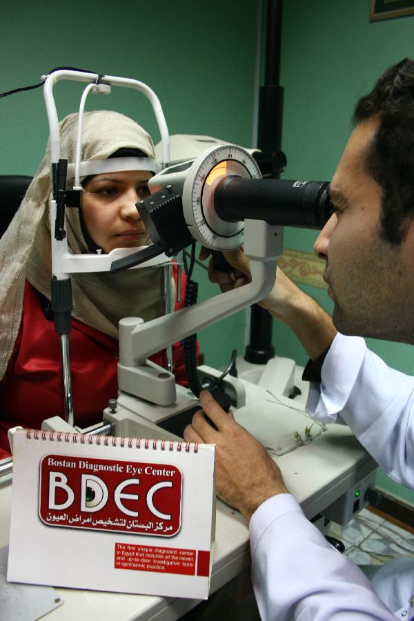
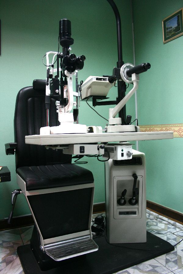
 Photo Slit lamp photography
The slit lamp exam uses an instrument that provides a magnified, three-dimensional (3-D) view of the different parts of the eye. During the exam, the doctor can look at the front parts of the eye, including the clear, outer covering (cornea), the lens, the colored part (iris), and the front section of the gel-like fluid (vitreous) that fills the large space in the middle of the eye.
Special lenses can be placed between the slit lamp and the cornea (or directly on the cornea) to view deeper structures of the eye, such as the optic nerve, retina and the area where fluid drains out of the eye (drainage angle). A camera may be attached to the slit lamp to take photographs of different parts of the eye.
Fluorescein dye eyedrops may be used during a slit lamp examination to make it easier to detect a foreign body, such as a metal fragment, or an infected or injured area on the cornea.
Why It Is Done
A slit lamp exam may be done:
- As part of a routine eye exam along with other procedures to evaluate the eye, such as ophthalmoscopy, vision testing or tonometry (to measure pressure in the eye).
- To look at structures in the back of the eye, such as the optic nerve or retina.
- To help detect disorders in the structures in the front of the eye, such as infection or injury to the cornea, cataracts, conjunctivitis or iritis.
- To help detect and monitor glaucoma or macular degeneration.
- To check for a foreign body, such as a metal fragment, on or in the eye.
- To detect eye problems that may be caused by other diseases, such as diabetes or rheumatoid arthritis. Routine slit lamp exams are important to detect eye problems at an early stage and to guide treatment if eye problems develop.
- To monitor complications such as bleeding after an eye injury.
- To monitor complications such as cataract formation that occur because of chemotherapy, radiation treatment or after a bone marrow transplant.
How To Prepare:
- If you wear glasses or contact lenses, you will need to remove them before the slit lamp examination.
- Eye drops may be used to widen (dilate) your pupils and to numb the surface of your eyes. Before the test, tell your doctor if you have glaucoma or are allergic to dilating or anesthetic eyedrops.
- If dilating drops are used, your eyes may be sensitive to light and you will have trouble focusing your eyes for several hours. If you know your eyes will be dilated, you may wish to arrange for someone to drive you home after the test. You also will need to wear sunglasses when you go outside or into a brightly lit room.
- Talk to your doctor about any concerns you have regarding the need for the test, its risks, how it will be done, or what the results will mean.
We provide slit lamp photography to document all the findings.
Confidentiality:
Information about our patients is collected in a variety of ways and for a variety of reasons (e.g. Providing care and treatment, managing and planning the center, training and educating staff, research etc.). It is stored on paper and on computerized systems.
Everyone working at the Bostan Diagnostic Eye Center has a legal duty to keep information about you confidential. Information will only ever be shared with people who have a genuine need for it (e.g. the medical staff or other professionals from whom you have been receiving care). Please be assured that we will treat your information with the highest degree of confidentiality.